TIME-RESOLVED DARK-FIELD X-RAY MICROSCOPY

Revealing Defect-Mediated Lattice Dynamics with Time-Resolved Dark-Field X-ray Microscopy

By combining pump–probe excitation with DFXM imaging of symmetry-breaking strain fields in the bulk, we visualize defect-mediated dynamics in Ge.DFXM experimental data and static · dynamic simulations support the observed contrast evolution and mode-conversion signatures.High X-ray penetration enables these dynamics to be imaged not only at the surface but throughout the bulk.

Seong Gook KimPOSTECH · Max Planck POSTECH/Korea Research Initiative

EXPERIMENTAL DATA & SIMULATIONS

Movies

01

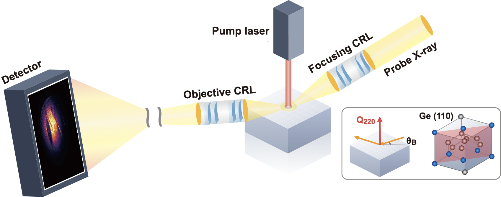
Overall pump–probe experiment

This movie summarizes the full time-resolved DFXM measurement sequence. Periodic intact-state measurements were interleaved with the pump–probe delay scans to verify reversible sample recovery and measurement reproducibility.
02–1

Representative case: annotated wave propagation

Blue arrows indicate the longitudinal strain wave propagating along the depth direction, while red arrows indicate the transverse elastic wave propagating in plane.

02–2

Larger-beam case: extended field of view

A larger beam size enables a broader sample region to be probed. Consistent longitudinal and transverse wave responses are also observed near the dislocation on the right; the red arrows specifically mark the transverse in-plane component.

02–3

Weak-contrast case: transverse wave also observed

The same in-plane transverse strain-wave response is observed at a weak-contrast dislocation from 1,000 to 6,000 ps. The red arrow marks its propagation.

03–1

Dynamic simulation: out-of-plane longitudinal component

A simplified COMSOL model with an idealized defect geometry shows that rapid thermoelastic expansion following normal-incidence laser excitation launches a longitudinal strain wave into the depth of the crystal.

03–2

Dynamic simulation: in-plane transverse component

The interaction between the wave and the dislocation induces mode conversion into an in-plane transverse component, visible as two linear wave fronts propagating in opposite directions.

Shown quantity: depth-direction displacement; therefore, the incident longitudinal wave is also visible in the XY plane.

SUPPLEMENTARY MATERIAL

Supplementary figures

Static and dynamic simulations supporting the observed DFXM contrast and strain-wave dynamics.

Ge dislocation geometries alongside their corresponding DFXM difference images
01 · DEFECT CONTRAST

Ge {111}⟨110⟩ dislocations

The figure compares different dislocation types with their corresponding DFXM difference images. In our experimental geometry, the contrast and morphology observed in DFXM depend on the dislocation type and orientation.

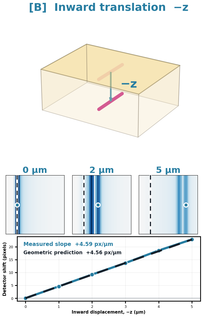
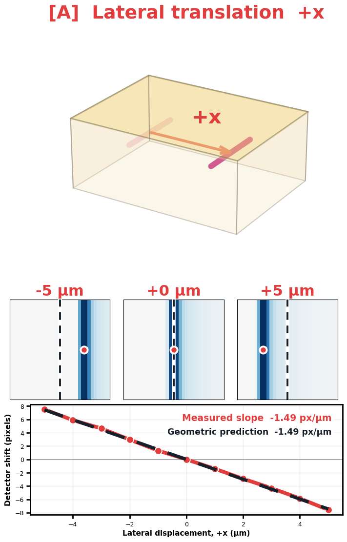
Simulated DFXM response to lateral translation of a localized strain field Simulated DFXM response to inward translation of a localized strain field
02 · DFXM SIMULATION

Simulated DFXM response to strain translation

To illustrate the DFXM signature of strain-wave propagation, a localized strain field was translated laterally and inward; the resulting contrast shifts are consistent with geometric predictions and do not imply physical defect motion.

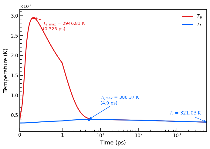
Calculated electron and lattice temperatures as a function of time
03 · TTM SIMULATION

Electronic and lattice temperature response

COMSOL two-temperature-model simulation of ultrafast laser excitation in germanium, showing rapid electron heating followed by electron–phonon energy transfer to the lattice.

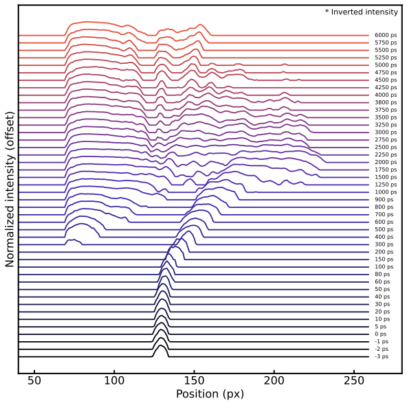
Stacked profiles annotated with in-plane and out-of-plane wave components and the initial defect position
04 · MODE CONVERSION

Defect-induced mode conversion

Stacked DFXM profiles show the emergence of an in-plane transverse component as the out-of-plane longitudinal strain wave interacts with a defect.